Strain-encoded cardiac magnetic resonance for the evaluation of chronic allograft vasculopathy in transplant recipients
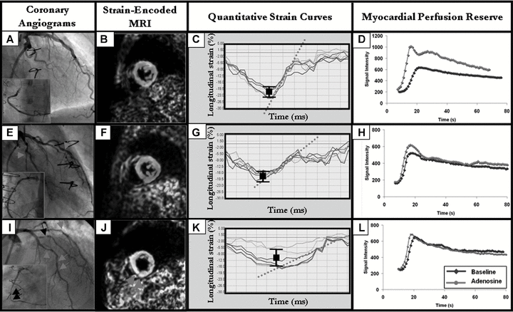
The aim of our study was to investigate the ability of Strain-Encoded magnetic resonance imaging (MRI) to detect cardiac allograft vasculopathy (CAV) in heart transplantation (HTx)-recipients. In consecutive subjects (n = 69), who underwent cardiac catheterization, MRI was performed for quantification of myocardial strain and perfusion reserve. Based on angiographic findings subjects were classified: group A including patients with normal vessels; group B, patients with stenosis <50%; and group C, patients with severe CAV (stenosis ≥ 50%). Significant correlations were observed between myocardial perfusion reserve with peak systolic strain (r = -0.53, p < 0.001) and with mean diastolic strain rate (r = 0.82, p < 0.001). Peak systolic strain and strain rate were significantly reduced only in group C, while mean diastolic strain rate and myocardial perfusion reserve were already reduced in group B and A. Myocardial perfusion reserve and mean diastolic strain rate had higher accuracy for the detection of CAV (AUC = 0.95, 95% CI = 0.87-0.99 and AUC = 0.93, 95% CI = 0.84-0.98, respectively) and followed peak systolic strain and strain rate (AUC = 0.80, 95% CI = 0.69-0.89 and AUC = 0.78, 95% CI = 0.67-0.87, respectively). Besides the quantification of myocardial perfusion, the estimation of the diastolic strain rate is a useful parameter for CAV assessment. In combination with the clinical evaluation, these parameters may be effective tools for the routine surveillance of HTx-recipients. © 2009 The American Society of Transplantation and the American Society of Transplant Surgeons.


